
Presentation
Fall from about 3m. No limb or chest injuries. Witnessed to hit head, confused.
Patient Data
Note: This case has been tagged as "legacy" as it no longer meets image preparation and/or other case publication guidelines.

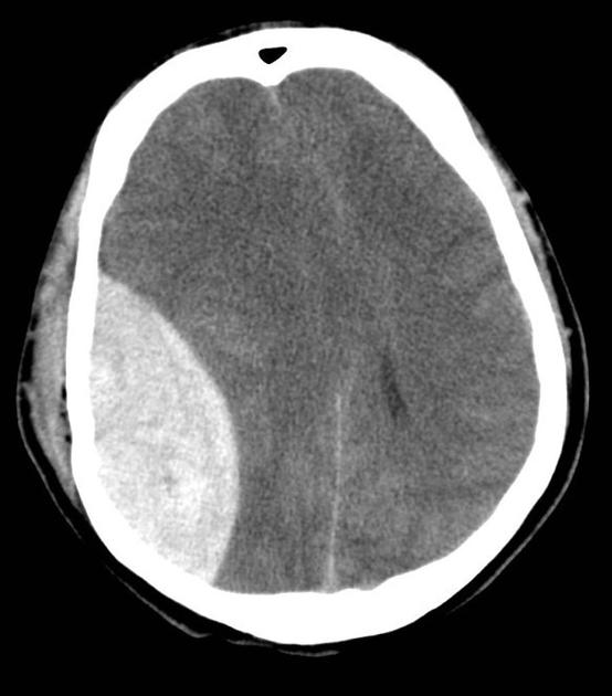
Large right-sided bi-convex (or lenticular) collection under the skull. It is hyperdense in comparison to surrounding brain tissue/parenchyma.
On this slice, there is midline shift with compression of the lateral ventricles.
There is also marked soft tissue swelling over the skull on the right side.
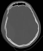
Using the bony window, there is a depressed, comminuted skull fracture overlying the area we know the collection is in.
Case Discussion
This case is a good example of extradural hemorrhage.
These usually occur after trauma (head injury). When the skull is fractured in high-energy injury, it can lead to tearing of the arteries sitting underneath the bone but above the dura.
It is important to get these bleeds quickly recognized and controlled, usually under direct neurosurgical care. There can be a "lucid interval" as the brains in these young patients can cope with the increased pressure from a bleed before deteriorating rapidly. There is also high risk of infection as these are usually open fractures.
CT is the method for imaging in these events as it is quick, widely available and can show bleeds and bone injuries well.
---
This case was contributed to Radiopaedia.org by Dr Sandeep Bhuta and the original can be viewed here. Note: patient presentation added to act as a teaching tool for EDH.


 Unable to process the form. Check for errors and try again.