
Presentation
3-month history of bilateral numbness in feet and thoracic back pain.
Patient Data


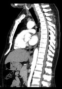
On contrast enhanced sequences, there is an enhancing soft tissue mass demonstrated in the posterior aspect of the spinal canal at the mid-thoracic level.
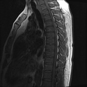
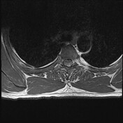
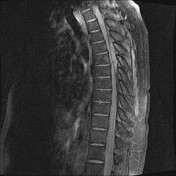
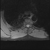

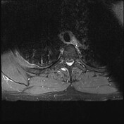
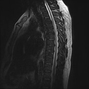
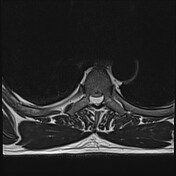
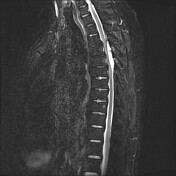
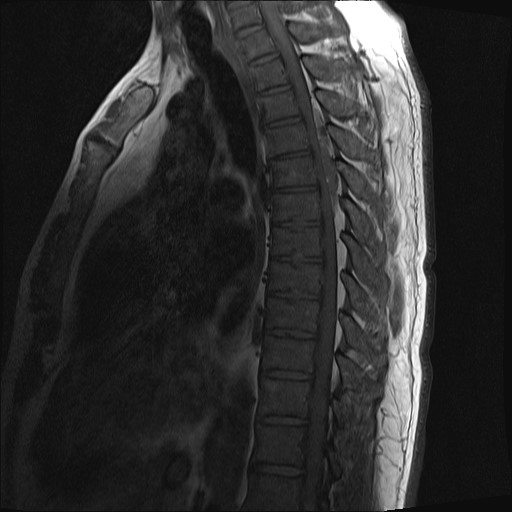
MRI of the thoracic spine demonstrates an extradural lesion posterior to the thoracic spinal cord. The well circumscribed and convex lesion exerts significant mass effect on the thoracic cord from T3 to T6/7 disc level. Anterior displacement of the dura is demonstrated. It is T1 hypointense and T2 iso- to hyperintense. There is uniform post-contrast enhancement. There is evidence of cord edema.
Histopathology report
MACROSCOPIC
Three specimens received.
1. Thoracic extradural lesion T3-T6 frozen section: One piece of pink tissue, 14x10x3 mm. 2 smears and 1 spreader, and one H&E prepared. 1A Frozen section remnant; 1B Serial sections remainder of tissue. Entirely submitted.
2. Thoracic extradural spinal lesion T3-6 MC&S: A piece of tan tissue, 8x6x5 mm. 2A Tissue bisected in toto.
3. Thoracic extradural spinal lesion T3-6 histo: 2x16x5 mm. 3A Bony tissue bisected (detail); 3B to 3F Serial sections tan tissue. Entirely submitted.
MICROSCOPIC
1 to 3. Thoracic extradural lesion T3-T6: The paraffin sections confirm the frozen section impression of a lesion composed of vessels, adipocytes and spindle cells. There are thin walled vessels of variable luminal size. Some are distended. No definite thrombi are identified within the vascular lumen. There are variably sized mature adipocytes with no significant cytological atypia. Some perivascular uniform spindle cells are noted. Mitotic figures are inconspicuous. Fragments of bone, bone marrow and tendinous tissue are included in specimen 3. There is no evidence of meningothelial whorls, necrosis or malignancy.
The spindle cells have the following immunohistochemical profile
Positive: ERG, CD34, CD31
Negative: GLUT-1, CK8/18, EMA, PR, desmin, S100, Melan A, HMB45, CD10 and PAX8.
SUMMARY
1 to 3. Thoracic extradural lesion T3-T6: Benign lesion composed of vessels and adipocytes
SUPPLEMENTARY REPORT
Thank you for the opportunity to review this biopsy with unusual morphology. It is a cavernoma composed of thick and thin-walled blood vessels as well as hemosiderin pigmentation. Some vascular spaces contain possible lymph. Presence of adipocytes amidst the vascular spaces in a cavernoma is uncommon but described in literature. There are no microthrombi within the vessels.
Impression: Cavernoma; no malignancy.
Case Discussion
The patient underwent spinal surgery during which the lesion was confirmed to be extradural and was noted to be easily removed from the dura. The histopathology results confirmed extradural spinal cavernous malformation.


 Unable to process the form. Check for errors and try again.