
Presentation
Hit on head.
Patient Data
Age: 50 years
Gender: Male
This case featured in our 2016 Trauma Radiology Course which is now available to view online.
From the case:
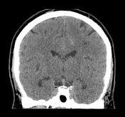
Falx subdural haematoma and sulcal subarachnoid haemorrhage


Download
Info
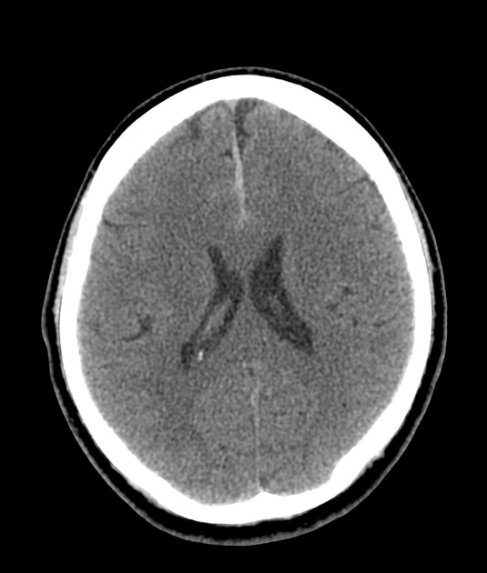
There is 7mm thickness acute falx subdural haematoma maximal in the frontal region. Low volume traumatic subarachnoid haemorrhage is seen within frontal sulci and callosal sulcus adjacent to the subdural, greater on the left. Small volume scalp haematoma at the vertex.
Case Discussion
Typical appearance of a thin traumatic falx subdural haematoma and low volume sulcal subarachnoid haemorrhage.


 Unable to process the form. Check for errors and try again.