
Presentation
Vietnamese. Ingestion of fresh water plants (Apium graveolens L, Enydra fluctuans L). Urticaria. Right upper quadrant pain.
Patient Data
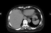

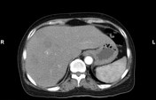
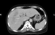
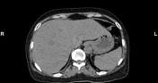
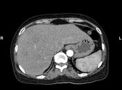
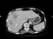
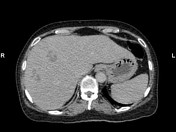

CT scan shows multiple, round, clustered, hypodense lesions, with peripheral contrast enhancement in the subcapsular liver
No bile duct dilatation
Left renal cyst
Blood test: WBC 7G/L. EO 1,87G/L. AST 40U/I. ALT 59U/I. GGT 25U/I
The patient had no ELISA test and no stool analysis for eggs
Case Discussion
This case has a differential diagnosis of pyogenic liver abscess, hepatic hydatid infection, malignancy, and fascioliasis.
These lesions demonstrated a clustered sign that can mimic pyogenic liver abscess, however, they are predominantly in the subcapsular liver and do not coalesce to form a large abscess cavity. And WBC no increased.
Hepatic hydatid infection doesn't have clustered sign, characterized by well-defined encapsulated cystic or multicystic masses. Daughter cysts, water-lily sign, and peripheral focal areas of calcification may be visualized.
The patient doesn't have any malignancy and necrotic metastases are usually not clustered or septated. No underlying cirrhosis.
In developing countries, parasites are the most common cause of hepatic abscesses. The patient usually ingested freshwater plants, containing larva(metacercaria) and blood test reveals eosinophilia. CT shows clustered, hypodense lesions, with peripheral contrast enhancement in the subcapsular liver. The history, symptoms, and imaging characteristics suggestive of fascioliasis
This patient was treated with Triclabendazole and was discharged.


 Unable to process the form. Check for errors and try again.