
Presentation
Left thigh pain.
Patient Data
Age: 50 years
Gender: Female
From the case:
Femoral diaphyseal stress fracture

Download
Info

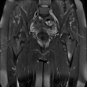
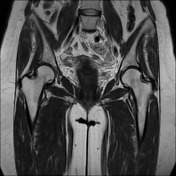
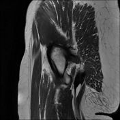
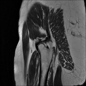
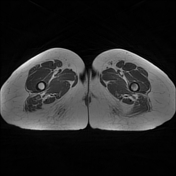
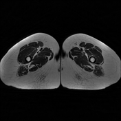
Left femoral upper shaft medial subcortical marrow edema signal with subtle small transversely oriented stress fracture line. Mild peri osseous soft tissue edema.
Findings are impressive of left femoral upper shaft medial stress fracture.

Download
Info
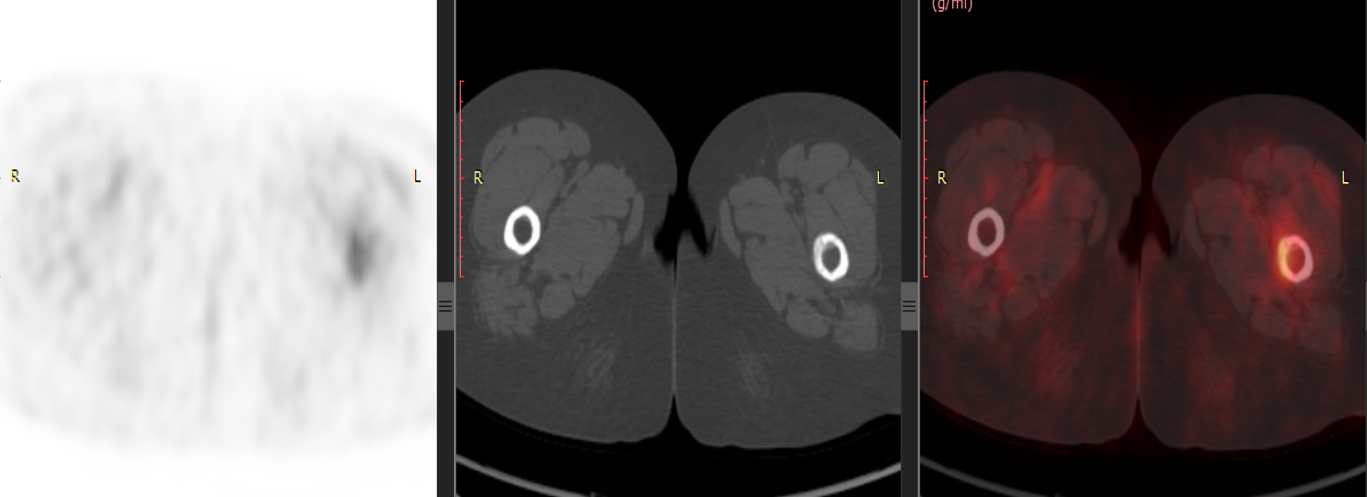
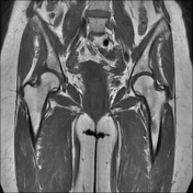
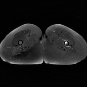
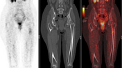
On PET-CT, the lesion shows mild increased FDG uptake with SUV max 2.4 with mild cortical thickening/sclerosis.
Case Discussion
MRI features of a stress fracture include periosteal reaction, endosteal bone marrow edema, and a linear osseous discontinuity.
On PET-CT, It usually shows mild increased FDG uptake.


 Unable to process the form. Check for errors and try again.