Presentation
G3L1. Anomaly scan at 20 wks normal. 27 wks scan - AFI - 20. This is 33 wks scan - AFI 25
Patient Data
Age: about 30 yrs
Gender: Female
From the case:
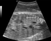
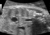
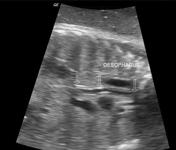
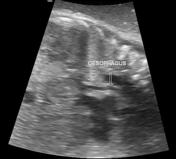
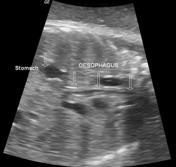
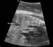
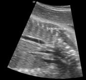
Fetal esophagus - dynamic scan in late onset polyhydramnios
![](https://prod-images-static.radiopaedia.org/images/5332630/428594d89c2bbcdefc50dfe811cdda_thumb.jpg)
![](https://prod-images-static.radiopaedia.org/images/5332638/a2c6a7629c43fce6b39e2e99ca7be4_thumb.jpg)
![](https://prod-images-static.radiopaedia.org/images/5332642/9f6e98ddd78f5324135f794df22b6d_thumb.jpg)
![](https://prod-images-static.radiopaedia.org/images/5332654/b8430564d99d788e2fe6e29207b4b0_thumb.jpg)
![](https://prod-images-static.radiopaedia.org/images/5332662/6e1d43b3258deb520a800f5b68a5c9_thumb.jpg)
![](https://prod-images-static.radiopaedia.org/images/5332663/d59213575b5c76f5287d0211cce38e_thumb.jpg)
![](https://prod-images-static.radiopaedia.org/images/5332675/69956fb91d36cd42dbe3bf1ee84437_thumb.jpg)
![](https://prod-images-static.radiopaedia.org/images/5332686/5f2bde5602f8b750305d80db734349_thumb.jpg)
Download
Info
![](https://prod-images-static.radiopaedia.org/images/5332630/428594d89c2bbcdefc50dfe811cdda_big_gallery.jpg)
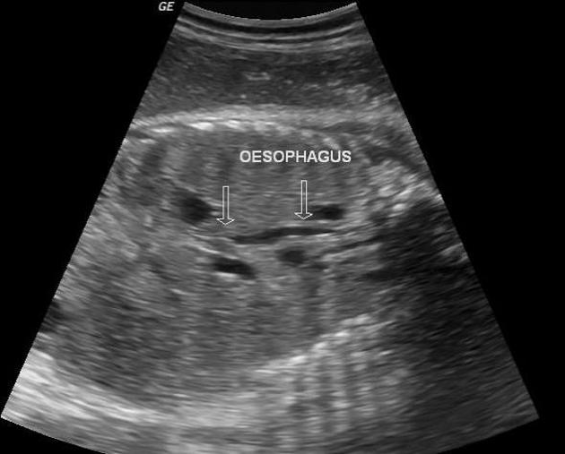
Multiple long axis images of fetal thoracic esophagus shows fluid filled and collapsed esophagus.
Case Discussion
Dynamic scan of esophagus at least rules out long segment atresia.
No cause for polyhydramnios is found at present.
Follow up.
- LSCS.
- No esophageal atresia.