
Presentation
Right mid-neck mass that appeared 2 weeks after birth. No trauma, fever or signs of infection.
Patient Data
Age: 3 weeks
Gender: Male
From the case:
Fibromatosis colli
Download
Info

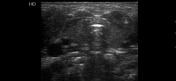
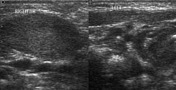
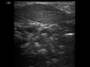
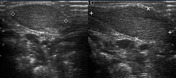
Ultrasound images show a fusiform enlargement of the right sternocleidomastoid muscle. It has a fibrillar pattern with no sign of masses or fluid nearby. No lymph nodes were found and the surrounding organs and tissues were normal. Left sternocleidomastoid muscle appeared normal.
Case Discussion
This case shows the typical features of fibromatosis colli. Unilateral, fusiform enlargement of sternocleidomastoid muscle. Contralateral sternocleidomastoid is normal.


 Unable to process the form. Check for errors and try again.