
Presentation
Post-traumatic left ankle pain.
Patient Data
Age: 15 years
Gender: Male
From the case:
Fibrous cortical defect

Download
Info
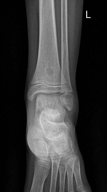
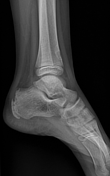
Well-defined geographic multiloculated, lucent lesion with a thin, sclerotic margin. It's eccentrically located in the metaphysis near the physis and presents a long axis parallel to the axis of the bone.
Case Discussion
Typical radiographic features of a fibrous cortical defect.


 Unable to process the form. Check for errors and try again.