
Presentation
Pain around left knee
Patient Data
Age: 16 years
Gender: Female
From the case:
Fibrous cortical defect

Download
Info
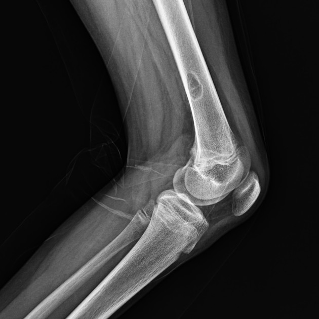
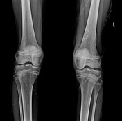
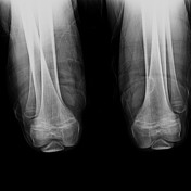
A cortical-based eccentric metadiaphyseal lucent lesion with a sclerotic rim is seen at the posteromedial aspect of the lower left femur. It shows overlying focal cortical thinning.
Case Discussion
Plain film features are characteristic of a fibrous cortical defect. It is a benign bony lesion that is usually small in size, occurs in skeletally immature children between age 2-15 years, and usually asymptomatic. It is typically seen in the distal femur, proximal and distal tibia.
It is one of the skeletal “Don’t touch” lesions. Lesions larger than 2 cm are called non-ossifying fibromas.


 Unable to process the form. Check for errors and try again.