
Presentation
Pain and locking sensation in knee
Patient Data
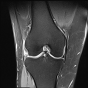

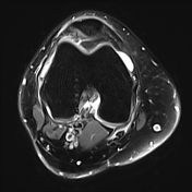

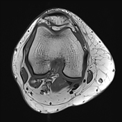
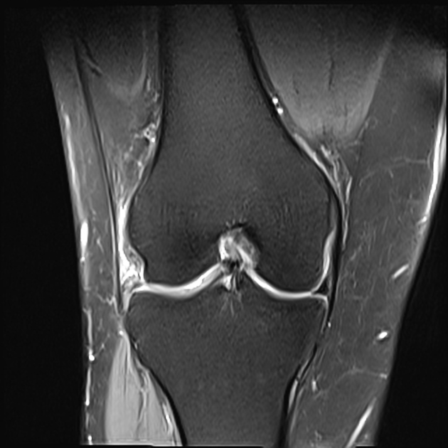
Tear of posterior horn and body of lateral meniscus is seen with displacement of posterior horn into intercondylar region and body at posterior aspect of truncated anterior horn.
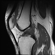
Medial and lateral collateral, anterior and posterior cruciate ligaments show maintained continuity.
Irregularity of articular cartilage is seen along lateral tibial condyle with subtle subchondral erosions and mild marrow edema.
Significant tendinosis of medial head of gastrocnemius tendon is seen.
Moderate to gross joint effusion is seen, extending to suprapatellar recess.
Enchondroma is seen in proximal fibula (incidental finding).
Case Discussion
Tear of posterior horn and body of lateral meniscus is seen with displacement of posterior horn into intercondylar region and body at posterior aspect of truncated anterior horn, suggestive of flipped meniscus.


 Unable to process the form. Check for errors and try again.