Presentation
Two liver lesions were noted as an incidental finding on CT performed as workup for night sweats.
Patient Data
![](https://prod-images-static.radiopaedia.org/images/8289524/26c92f71b3b853d573bbc9b861e527_thumb.jpg)
![This study is a stack](/packs/stack-YQKLCKBI.gif)
![](https://prod-images-static.radiopaedia.org/images/8289707/6b494baba379d80b387baebcb36883_thumb.jpg)
![This study is a stack](/packs/stack-YQKLCKBI.gif)
![](https://prod-images-static.radiopaedia.org/images/8289775/b458923daf75231fe18be21ec421e2_thumb.jpg)
![This study is a stack](/packs/stack-YQKLCKBI.gif)
![](https://prod-images-static.radiopaedia.org/images/8289873/8df8827db273b54219ad86e15493e9_thumb.jpg)
![This study is a stack](/packs/stack-YQKLCKBI.gif)
![](https://prod-images-static.radiopaedia.org/images/8289809/2ae52931e977736340b97a62af653a_thumb.jpg)
![This study is a stack](/packs/stack-YQKLCKBI.gif)
![](https://prod-images-static.radiopaedia.org/images/8289843/3641a9f2a3039904b920117bb5120a_thumb.jpg)
![This study is a stack](/packs/stack-YQKLCKBI.gif)
![](https://prod-images-static.radiopaedia.org/images/8289938/313a88f4df3e894fe2ab302e6e8bdc_thumb.jpg)
![This study is a stack](/packs/stack-YQKLCKBI.gif)
![](https://prod-images-static.radiopaedia.org/images/8289616/040b046122aeac06ec7d6349944a31_thumb.jpg)
![This study is a stack](/packs/stack-YQKLCKBI.gif)
![](https://prod-images-static.radiopaedia.org/images/8290042/124519dd16020e32e6dd670abd8165_thumb.jpg)
![This study is a stack](/packs/stack-YQKLCKBI.gif)
![](https://prod-images-static.radiopaedia.org/images/8290146/5c092a5a625dc2aa76520b83ccd473_thumb.jpg)
![This study is a stack](/packs/stack-YQKLCKBI.gif)
![](https://prod-images-static.radiopaedia.org/images/8290250/9c5e4e836400daded01c7790d24747_thumb.jpg)
![This study is a stack](/packs/stack-YQKLCKBI.gif)
![](https://prod-images-static.radiopaedia.org/images/8290416/00a2f82f9a2cac80f5e3ef38b261aa_thumb.jpg)
![This study is a stack](/packs/stack-YQKLCKBI.gif)
![](https://prod-images-static.radiopaedia.org/images/8289524/26c92f71b3b853d573bbc9b861e527_big_gallery.jpg)
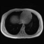
There is no evidence of liver parenchymal disease.
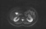
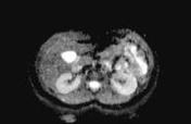
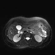
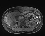
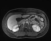
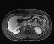
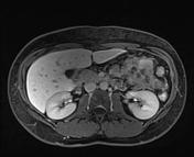
2 focal liver lesions are identified:
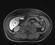
- In segment 5/6, there is a 2.9 centimeter rounded lesion which is minimally T2 hyperintense and has a relatively T2 hyperintense central component, which is T1 hypointense. It does not contain fat. It does not restrict diffusion. There is vivid arterial enhancement without washout. The central component does not enhance. The lesion however concentrates Primovist.
- In segment 3, there is a 2.1 centimeter rounded lesion which is minimally T2 hyperintense. It does not contain fat. It does not restrict diffusion. There is vivid arterial enhancement without washout. A central component does not enhance. The lesion however concentrates Primovist at the periphery.
There is a tiny focus seen in the subcapsular region of segment IVa anteriorly, only visible on the arterial phase, with unknown etiology and significance.
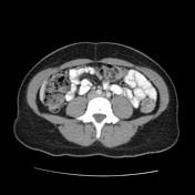
The biliary tree is non-dilated. The pancreas, kidneys, adrenal glands, spleen are normal in appearance. No free fluid or lymphadenopathy.
Conclusion: Two focal liver lesions with MR appearances consistent with focal nodular hyperplasia.
![](https://prod-images-static.radiopaedia.org/images/8290471/bb903e73361b522f364c3206febb9a_thumb.jpg)
![This study is a stack](/packs/stack-YQKLCKBI.gif)
![](https://prod-images-static.radiopaedia.org/images/8290601/b93a72189f9091fe4b949776cd3280_thumb.jpg)
![This study is a stack](/packs/stack-YQKLCKBI.gif)
![](https://prod-images-static.radiopaedia.org/images/8290711/c48daf7a7efab93e1649ffc9b547aa_thumb.jpg)
![This study is a stack](/packs/stack-YQKLCKBI.gif)
![](https://prod-images-static.radiopaedia.org/images/8290536/2e0a9c7751e2eee5309d66023d2383_thumb.jpg)
![This study is a stack](/packs/stack-YQKLCKBI.gif)
![](https://prod-images-static.radiopaedia.org/images/8290647/85016bf77d18fb3747f83dae989de4_thumb.jpg)
![This study is a stack](/packs/stack-YQKLCKBI.gif)
![](https://prod-images-static.radiopaedia.org/images/8290471/bb903e73361b522f364c3206febb9a_big_gallery.jpg)
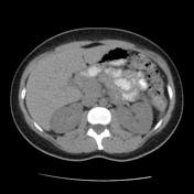
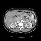
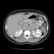
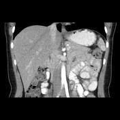
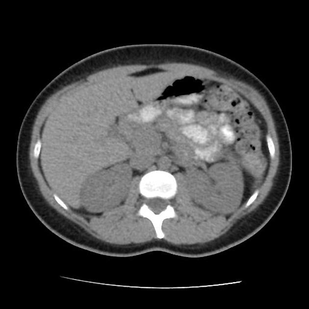
Two enhancing lesions in segment 6 and 3 with central hypodensity (nonenhancing) and no washout.
Case Discussion
Typical appearances of focal nodular hyperplasia, one of the few entities that can hyperconcentrate hepatocyte specific contrast agent.