
Presentation
Tender swelling of the anterior aspect of the mid-left leg in a military patient. No clear history of trauma.
Patient Data
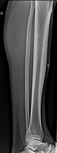
A radio-opaque foreign body is seen in the soft tissue of the anterior aspect of the mid-tibial shaft with thickening of the surrounding subcutaneous fatty tissue. No underlying bone lesion is seen.
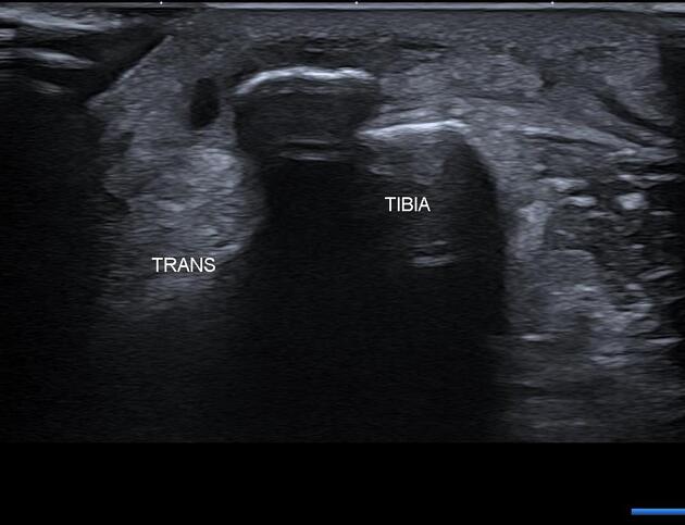
On ultrasound images, the foreign body appears as an arciform echogenic structure with posterior acoustic shadowing and heterogeneous hypoechoic surrounding soft tissue as well as thickening with increased echogenicity of the subcutaneous fatty tissue.
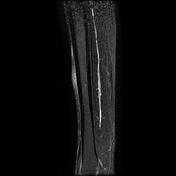

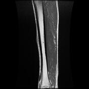

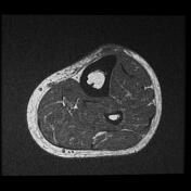
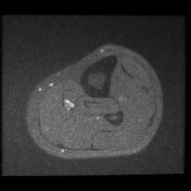
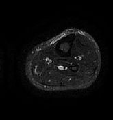
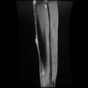

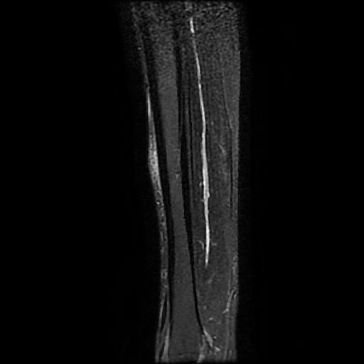
On the MRI sequences, the foreign body is of low signal on all sequences with enhancement of the surrounding inflammatory granuloma as well as thickening with enhancement of the subcutaneous fatty tissue. The underlying tibial cortex is intact with no bone marrow edema.
Case Discussion
This case illustrates the radiographic, ultrasound and MRI appearance of a foreign body granuloma.
Foreign body granuloma is an inflammatory tissue reaction around retained foreign bodies after penetrating trauma. Sometimes they can be mistaken for soft tissue tumors when the foreign body is not seen or recognized and the symptoms are non-specific or there is no clear history of trauma.


 Unable to process the form. Check for errors and try again.