
Presentation
Right proptosis. History of right sinonasal surgery.
Patient Data
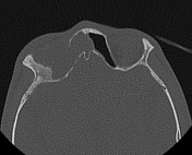


Enlarged right orbit with rarified superior orbital ridge and upper part of its medial wall.
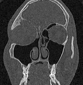
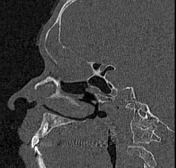
Expanded right frontal sinus by homogeneous density with smooth scalloped bony wall and dilated frontal outflow tract.
Right maxillary antrostomy, middle turbinectomy and ethmoidectomy.

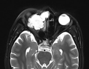
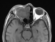
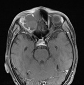
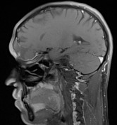

Totally opacified and expanded right frontal sinus with expansile thinning of its bony walls, up to complete absence of its floor (right orbital roof).
The right orbital extension measures 2x3 cm. It exerts stretching and indentation of the superior muscle complex, as well as the globe, causing right proptosis.
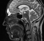
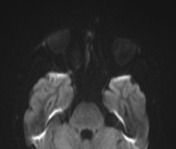
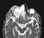
The overall sinus shows marked T2 hyperintensity, low-to-intermediate T1 signal, and thin peripheral rim of mucosal enhancement. No solid enhancing nodules. Clear fat planes. Other sinuses are aerated.
Right maxillary antrostomy, middle turbinectomy and ethmoidectomy.
Case Discussion
Features are consistent with right frontal mucocele. The frontal sinus is the most common site of paranasal sinus mucocele, followed by the ethmoid sinuses.
It occurs as a result of obstruction of the ostium of the sinus, and can be due to sequels of previous sinonasal surgery as it occurs at the same side of the previous surgery.


 Unable to process the form. Check for errors and try again.