
Presentation
Headache.
Patient Data
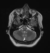

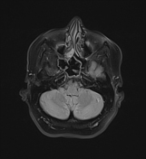
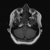
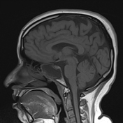
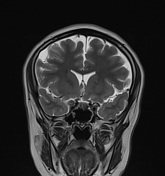

Expanded right sphenoid sinus elicits intermediate signal in T1WI. In T2WI, the sinus elicits low signal intensity with minimal peripheral hyperintense mucosal thickening.
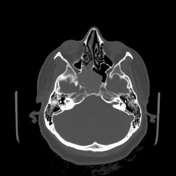
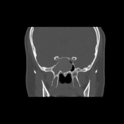
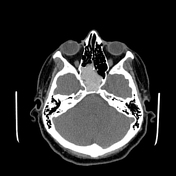
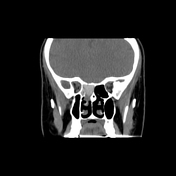

Almost complete opacification of the right sphenoid sinus with internal hyperdense contents associates with smooth expansion and thinning out of bony sinus walls yet no gross bony destruction seen. It is encroaching upon the left sphenoid sinus and right posterior ethmoid air cells.
Case Discussion
Here is a case of right sphenoid fungal sinusitis with expansion suggesting mucocele. The hypointense T2 signal may be mistaken for a normally aerated sinus (pseudo-pneumatized). T1 signal changes and a T2 hyperintense peripheral inflamed mucosal thickness are helpful to avoid a missed diagnosis.
The low T2 signal or signal void is due to the high concentration of various metals such as iron, magnesium, and manganese concentrated by fungal organisms as well as high protein and low free water content in allergic mucin.


 Unable to process the form. Check for errors and try again.