
Presentation
Pain and deformity after a fall.
Patient Data
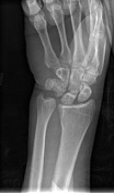
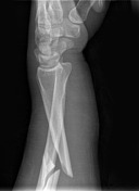

There is a comminuted fracture of the middle third of the radius, demonstrating approximately 20 degrees of posterior angulation with a butterfly fragment posteriorly at the fracture site. The ulnar head is dislocated anteriorly, with widening of the distal radio-ulnar joint. The scapholunate joint space is borderline widened.

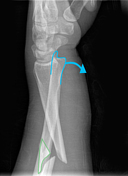

The pathological changes seen in this fracture-dislocation injury pattern are outlined.
DP:
- The widened distal radioulnar joint is indicated by the blue line
- The loss of the normal contour between the radius and ulna because of the dislocated ulnar head is indicated by the green line
- The borderline widened scapholunate joint spaces are indicated by the yellow line (should be <3.5mm)
Lateral:
- The anteriorly dislocated ulnar head is outlined in blue
- The comminuted butterfly fragment is outlined in green
Case Discussion
It is critical to identify the dislocated ulnar head in this case, as it changes it from a simple radius fracture to a Galeazzi injury pattern, which greatly changes the patient management and outcomes as this is an unstable injury and the extent of injury to other structures is much greater. Anterior dislocation of the ulna (as in this case) is less common than posterior dislocation.


 Unable to process the form. Check for errors and try again.