
From the case:
Gallbladder polyp
Download
Info

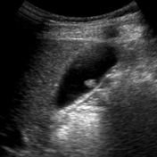
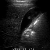
Two selected ultrasound images demonstrate a pedunculated 7mm rounded mass projecting into the lumen of the gallbladder. It is of intermediate echogenicity and demonstrates no posterior acoustic shadowing.
Features are those of a gallbladder polyp. Given its size and appearances, follow-up is necessary to ensure no growth.


 Unable to process the form. Check for errors and try again.