
Presentation
Incidental finding of a gallbladder lesion on ultrasound.
Patient Data

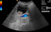
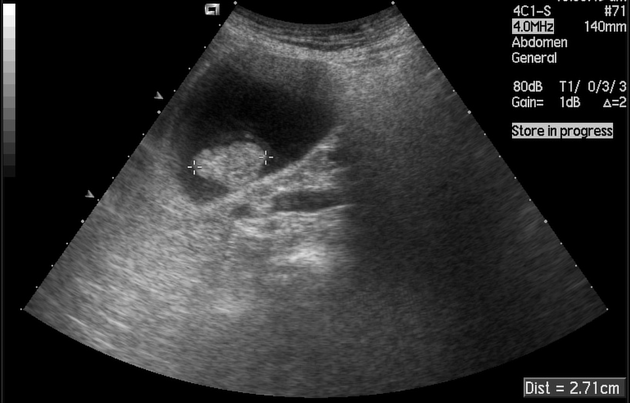
There is a 27 mm polypoid lesion within the lumen of the gallbladder that is fixed to the wall. There is no posterior acoustic enhancement or ring-down artefact. There is internal vascularity on Doppler imaging. No extramural extension beyond the gallbladder wall.
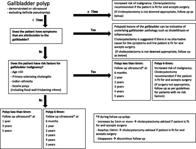
This chart is a summary of the 2017 joint European guidelines.
Source: Wiles, R., Thoeni, R.F., Barbu, S.T. et al. Eur Radiol (2017). doi:10.1007/s00330-017-4742-y
Licence: Creative Commons Attribution 4.0 International License.
Case Discussion
The management and follow-up of gallbladder polyps is controversial and practice varies between centers. However, due to the increased risk of malignancy, polyps measuring 10mm or greater should be considered for cholecystectomy 1.
This patient underwent cholecystectomy and a gallbladder adenoma with low-grade dysplasia was confirmed by histopathology.
Case courtesy of Dr Jamie Franklin and Dr Helen Bungay on behalf of the British Society of Gastrointestinal and Abdominal Radiology.


 Unable to process the form. Check for errors and try again.