
Presentation
Recurent upper right quadrant pain. Ultrasound showed a hyperechoic intraluminal, sessile lesion of the gallbladder without acoustic shadowing.
Patient Data
Age: 50 years
From the case:
Gallbladder polyp - CT

Download
Info
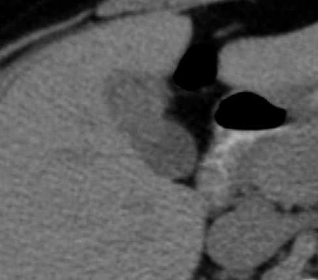
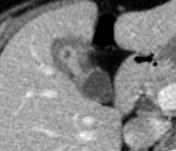
Within the lumen of the corpus and fundus of the gallbladder, a soft tissue attenuating lesion could be found, which demonstrated enhancement after IV contrast.
Case Discussion
MSCT showed a sessile gallbladder polyp of about 15 mm, that enhanced avidly with contrast.


 Unable to process the form. Check for errors and try again.