
Presentation
Hematuria.
Patient Data
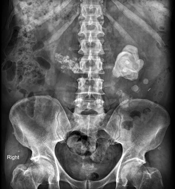
Multiple variable-sized hyperdense foci compatible with urinary tract calculi indicate an unusually medial position of the lower poles of both kidneys.


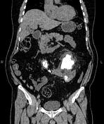
The lower poles of both kidneys are seen pointing medially and fused across the midline by an isthmus of functioning renal tissue, with anteriorly malrotated hila denoting horseshoe kidney.
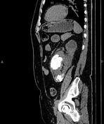
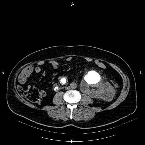
A 70 mm huge stone is noted in the left renal collecting system with resultant severe hydronephrosis and renal parenchymal atrophy. Smaller stones are also evident in the lower part of the left kidney.
Additionally, multiple adjacent small stones and some parenchymal calcification foci are observed in the lower pole of the right kidney. However, the upper pole of the right kidney shows normal parenchymal thickness without hydronephrosis. The right ureter is also normal.
Several small air bubbles are noted in the right renal collecting system and urinary bladder, probably due to recent catheterization.
The prostate gland is relatively enlarged.
Case Discussion
Poor urinary drainage can cause complications in horseshoe kidney. Hydronephrosis may be secondary to pelviureteric junction obstruction, and nephrolithiasis can be noted in up to 60% of patients. There is also an increased risk of infection and pyeloureteritis cystica, renal malignancy, and renovascular hypertension.


 Unable to process the form. Check for errors and try again.