
Presentation
Abdominal pain with constitutional symptoms for 2 months. An ultrasound of the abdomen revealed a large cystic mass of unknown origin.
Patient Data


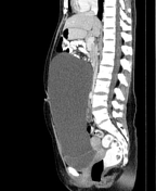
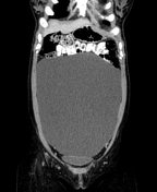

Large, thin-walled cystic mass (average density of +1 to +7 HU) occupying the mid to lower abdomen. There are no enhanced solid components or septations within. No intralesional fat or calcifications. Both right and left ovaries are visualized; however, there is no clear plane with this mass. It causes displacement of the small bowels superiorly and compression of the urinary bladder inferiorly. No bowel dilatation. No hydronephrosis or hydroureter.
The uterus is anteverted, with mild hydrometra within. Douglas' pouch contains very little fluid.
Tiny hypodense lesion in segment VII of the liver. The rest of the solid organs are preserved. There is no abdominal or pelvic lymphadenopathy.
Case Discussion
It is often difficult to confidently ascertain the origin of large masses, as in this case. Differential diagnoses to consider includes an ovarian cyst (no clear plane with the ovaries), a mesenteric or omental cyst, or a duplication cyst.
Intraoperatively, this large cystic mass was found to be arising from the left ovary.
Histopathology
Macroscopic description: The specimen consists of collapsed cystic tissue with an ovary seen within the cyst. Omental specimen containing yellowish tissue.
Microscopic description: Sections of the cyst show ovarian and fibro-collagenous stroma lined by stratified ciliated columnar epithelium forming broad branching papillae. No marked cellular atypia or stromal invasion was seen. Fallopian tubes are unremarkable. Omental specimens showed sheets of congested mature adipocytes with focal lymphoid aggregates. No malignancy was seen.
Diagnosis: Serous cystadenoma of the ovary. No malignancy was seen.


 Unable to process the form. Check for errors and try again.