
Presentation
Palpable mass in the back of the knee initially mistaken for a popliteal cyst.
Patient Data
Age: 65 years
Gender: Male
From the case:
Giant popliteal artery aneurysm


Download
Info
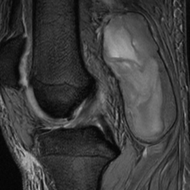
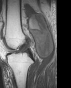
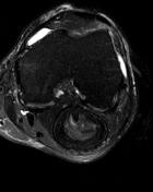
Large oval, sharply delineated, popliteal mass in continuation with the popliteal artery. Hyperintense signal on T1. Hypo-, iso-, and hyperintense signals with concentric layering on T2. Compression of the popliteal vein.
Case Discussion
Typical MRI aspect of a large thrombosed aneurysm. A popliteal cyst can be ruled out easily by identifying the gastrocnemius semimembranosus bursa in the medial popliteal fossa, which contains only minimal fluid.


 Unable to process the form. Check for errors and try again.