
Presentation
Right-sided abdominal pain and palpable mass. She had mildly elevated CRP, otherwise lab results were normal. Patient had no other symptoms.
Patient Data
Age: 25 years
Gender: Female
From the case:
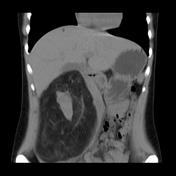
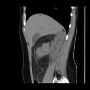
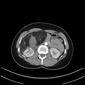
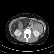
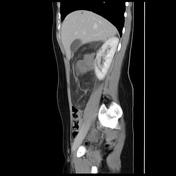
Giant renal angiomyolipoma


Download
Info
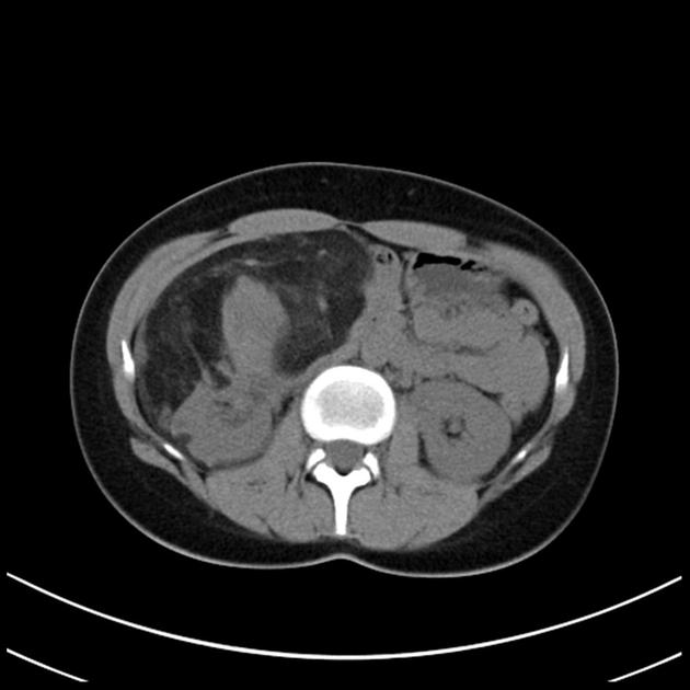
Mixed fat-solid vascularized giant mass in the renal space with central foci measuring around 40 HU - probable hematoma. Note additional small fat foci in both kidneys and one small focus in the liver.
Contributor: Dr Mitja Četina
Case Discussion
Mass and right kidney were removed.
Histology report (summary)
Angiomyolipoma growing out of the renal cortex, tumor cells were mildly pleomorphic, without mitosis or necrosis. MDM-2 negative. Additional small foci of angiomyolipomas in the kidney.


 Unable to process the form. Check for errors and try again.