
Presentation
Asymptomatic patient for routine screening mammogram.
Patient Data

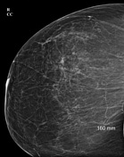
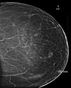
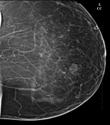
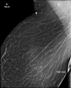
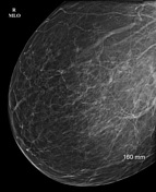

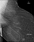
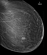
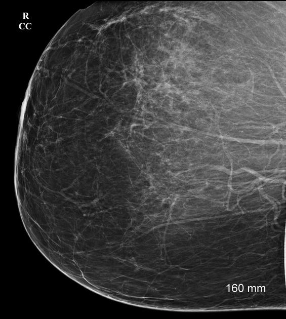
Large breasts with predominantly fatty parenchymal density. A benign looking lobular soft tissue density is seen in the lower inner quadrant of the left breast. No architectural distortion, pleomorphic microcalcification clusters, abnormal skin thickening or nipple retraction is seen in either breast. No significant axillary lymphadenopathy is seen.
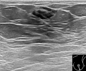
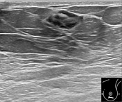
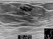

A well-defined ovoid-shaped mildly heterogenous hypoechoic lesion measuring 8 x 12 mm is noted at 6 0'clock position in the superficial left breast. No internal vascularity is seen in it on color Doppler ultrasound examination.
Case Discussion
Patient is morbidly obese (height=169 cm, weight=144 kg, BMI=50.42 kg/m2). Based on the history, mammographic findings are suggestive of gigantomastia/macromastia (type 1a). Left breast nodule is a BI-RADS 3 lesion.
-
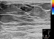
Left breast nodule was biopsied under ultrasound-guidance and histopathology showed benign atrophic lobules in loose collagenous connective tissue stroma. No evidence of malignancy noted.


 Unable to process the form. Check for errors and try again.