
Glenolabral articular disruption (GLAD lesion) with Buford complex and supraspinatus tear
Updates to Case Attributes
1. Episode/sThis patient with a history of prior anterior glenohumeral instability with recurrent shoulder dislocations demonstrates an anatomical variant that is the Buford complex and glenolabral articular disruption (GLAD) lesion) with an intra-rticular chondral fragment. There is no anteroinferior glenoid bone loss and the Hill-Sachs defect (onis moderate-sized making this an on-track lesion). Significant chondral loss with In addition, there is a fragment displaced into the axillary recess. Buford complex noted.
2. Full thickness partial width anterior supraspinatus footplate tear extendingtear, which is important to note as a delamination tear into the critical zonerotator cuff repair after shoulder dislocation may lead to better outcomes.
References changed:
- Gombera M, Gomberawalla M, Sekiya J. Rotator Cuff Tear and Glenohumeral Instability : A Systematic Review. Clin Orthop Relat Res. 2014;472(8):2448-56. <a href="https://doi.org/10.1007/s11999-013-3290-2">doi:10.1007/s11999-013-3290-2</a> - <a href="https://www.ncbi.nlm.nih.gov/pubmed/24043432">Pubmed</a>
Updates to Link Attributes
Updates to Link Attributes
Updates to Primarylink Attributes
Updates to Study Attributes
Image MRI (T1) ( update )
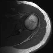
Image 1 MRI (PD fat sat) ( update )
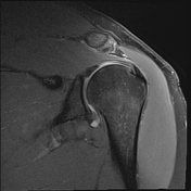
Image 2 MRI (PD) ( update )
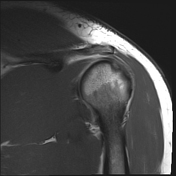
Image 3 MRI (PD fat sat) ( update )
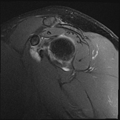
Image 4 MRI (PD) ( update )
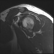
Image 5 MRI (PD fat sat) ( update )
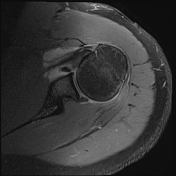
Image 6 MRI (T1) ( update )


 Unable to process the form. Check for errors and try again.