
Presentation
Seizures and right-sided hemiplegia.
Patient Data
Age: 50 years
Gender: Male
From the case:
Glioblastoma IDH wild-type


Download
Info

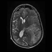
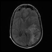
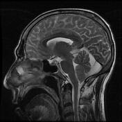
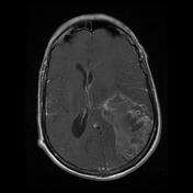
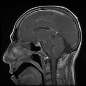
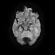
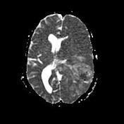
MRI demonstrates a large heterogenous signal intra-axial mass in the left parietal-temporal lobes. It exerts a significant mass effect with a midline shift and compression of the left lateral ventricle occipital horn and is surrounded by vasogenic edema.
The lesion shows patches of diffusion restriction and heterogenous post-contrast enhancement.
Case Discussion
MRI features are most consistent with a high-grade (WHO grade IV) primary glial tumor. Biopsy confirmed this to be a glioblastoma multiform, IDH wild type, WHO grade IV.
IHC results:
Oligo2: positive
GFAP: positive
IDH1: negative
EMA: negative


 Unable to process the form. Check for errors and try again.