
Presentation
Two brothers with the same condition.
Patient Data


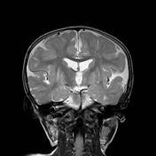
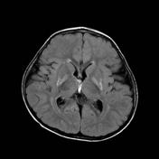
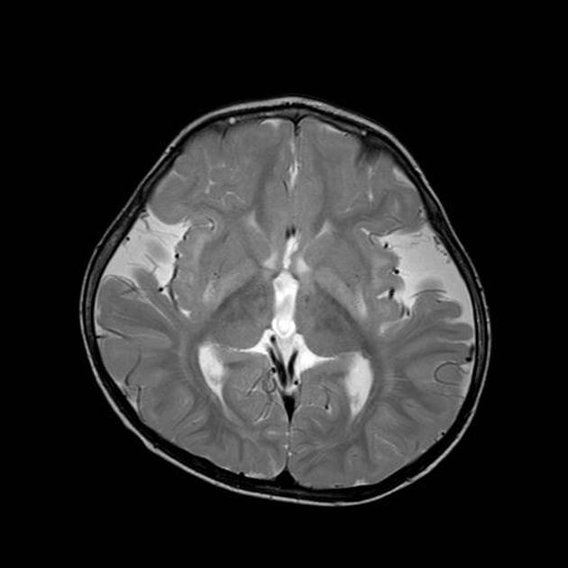
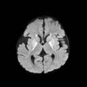
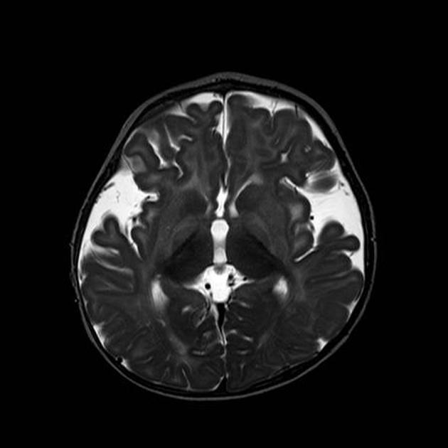
There is abnormal high T2/FLAIR in the putamina and globus pallidi, which also demonstrate volume loss and restricted diffusion. Further signal abnormality is seen in the tegmental tracts along the floor of the fourth ventricles.
Wide CSF spaces are seen anterior to the temporal poles, and within the sylvian fissures which appear under-opercularised.

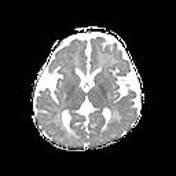
Younger brother scanned at 8 mth of age
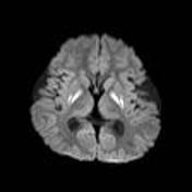
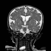
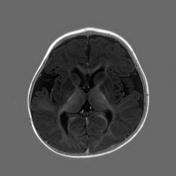
The findings are similar to his older brother, but note that there is less volume loss of the putamina and globus pallidi. Also, no tegmental abnormality is seen at this stage.
Wide CSF spaces anterior to the temporal poles and in the Sylvian fissures are again seen, but in this case, the prominence of extra-axial CSF spaces over the convexity is better appreciated.
Case Discussion
Both brothers are affected by glutaric aciduria type 1. It is inherited in an autosomal recessive pattern, and their parents are first degree cousins. Although the findings described above are not by themselves specific, the combination of findings is highly suggestive of the diagnosis1. Macrocephaly is also a feature of this condition, and although it is difficult to appreciate macrocephaly on MRI, head circumference should be sought from the referring clinicians, as it significantly limits the differential2.
In the case of the older brother, imaging suggested the diagnosis before any metabolic investigations were carried out. Diagnosis was subsequently confirmed by urinary analysis and DNA sampling. His younger brother was screened at birth, allowing early post-natal diagnosis.
The changes in the lentiform nuclei are related to acute metabolic crises, and are a major cause of morbidity3. The more pronounced changes in the older brother reflect the longer duration of illness. Early treatment with a low protein diet may help slow neurological deterioration4, but in this family, treatment was hampered by language barrier and questionable compliance.
Importantly, glutaric aciduria type 1 can present with sub-dural haematomas as the increased extra-axial sub-arachoid spaces means the coursing bridging veins can easily rupture. This should not be mistaken for non-accidental injury.


 Unable to process the form. Check for errors and try again.