Presentation
Left lower abdominal pain in History of cesarean section 3 months prior.
Patient Data
![](https://prod-images-static.radiopaedia.org/images/63573685/8079bf7498a7b5bd36a51b2ce86ebbaec1680cf90b7ce36842b979f2976d537b_thumb.jpeg)
![This study is a stack](/packs/stack-YQKLCKBI.gif)
![](https://prod-images-static.radiopaedia.org/images/63573709/3b8ed88b1c6280362cd3e1fb78ccfe8ff9bf784165ea13bffbc6d5e6dbe292f8_thumb.jpeg)
![This study is a stack](/packs/stack-YQKLCKBI.gif)
![](https://prod-images-static.radiopaedia.org/images/63573743/ccd0369b1104950c1430d329f6cbc4275eaf55f2f814c81ab11f63a939a86ac4_thumb.jpeg)
![This study is a stack](/packs/stack-YQKLCKBI.gif)
![](https://prod-images-static.radiopaedia.org/images/63573783/1e7a4f4682ebe0cdc5c461ae9cb3790ed9a7fa9264c8f67dafb08465a537064a_thumb.jpeg)
![This study is a stack](/packs/stack-YQKLCKBI.gif)
![](https://prod-images-static.radiopaedia.org/images/63573812/1dc4767c5d6408eac75ebde262eb9ec071aae80f151545419b85dabfd35ac7f3_thumb.jpeg)
![This study is a stack](/packs/stack-YQKLCKBI.gif)
![](https://prod-images-static.radiopaedia.org/images/63573855/d234a35310001064032c46769a8b57a1ae2ca60a4d2f829818a2ab68cfa99372_thumb.jpeg)
![This study is a stack](/packs/stack-YQKLCKBI.gif)
![](https://prod-images-static.radiopaedia.org/images/63573873/5d4e02e2fa047178cf34f4d3d0d4fbe5c25d1882d857a661890d291c8a860122_thumb.jpeg)
![This study is a stack](/packs/stack-YQKLCKBI.gif)
![](https://prod-images-static.radiopaedia.org/images/63573685/8079bf7498a7b5bd36a51b2ce86ebbaec1680cf90b7ce36842b979f2976d537b_big_gallery.jpeg)
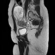
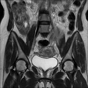
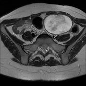
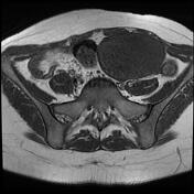
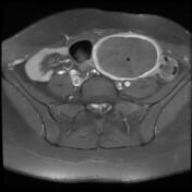
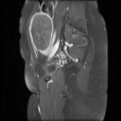
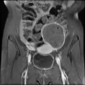
Large well-circumscribed mass in the right upper pelvic region of a low signal on T1 and high signal on T2, containing serpiginous structures of intermediate to low signal with peripheral enhancement on postcontrast sequences. No pelvic lymphadenopathy or free fluid in the peritoneal cavity.
![](https://prod-images-static.radiopaedia.org/images/63573894/cc42d8d524b9a360a4d64e1637c64cfa85c70566800871bf76da83d02f173d65_big_gallery.jpeg)
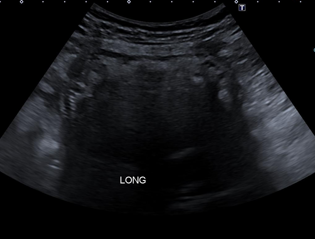
The ultrasound images demonstrate a hypoechoic fluid collection containing an arciform echogenic structure with intense posterior acoustic, highly suggestive of gossypiboma
Case Discussion
The clinical history, ultrasound and MRI features are most consistent with a gossypiboma that was confirmed at surgery (laparoscopic approach) as a surgical gauze.