
Presentation
3 weeks of headaches.
Patient Data
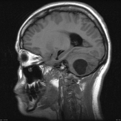

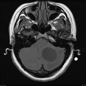
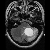
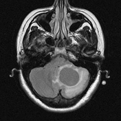
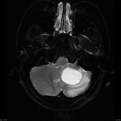
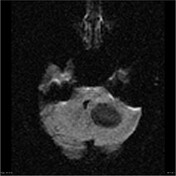

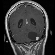

Left intra-axial posterior fossa mass is characterized by a large cyst with enhancing nodule The nodule abuts pia inferiorly and a small superficial vessel is seen entering the nodule (best seen on T2 weighted images). No evidence of abnormal susceptibility or diffusion restriction.
Associated surrounding edema and mass effect causes effacement of the adjacent sulci and compression of the fourth ventricle. Dilated lateral ventricles and third ventricle with transependymal edema present consistent with hydrocephalus. Tonsillar herniation, presumed secondary to mass effect from the tumor is also noted.
No further lesions are identified.
The patient went on to have a posterior fossa craniotomy and the tumor resected.
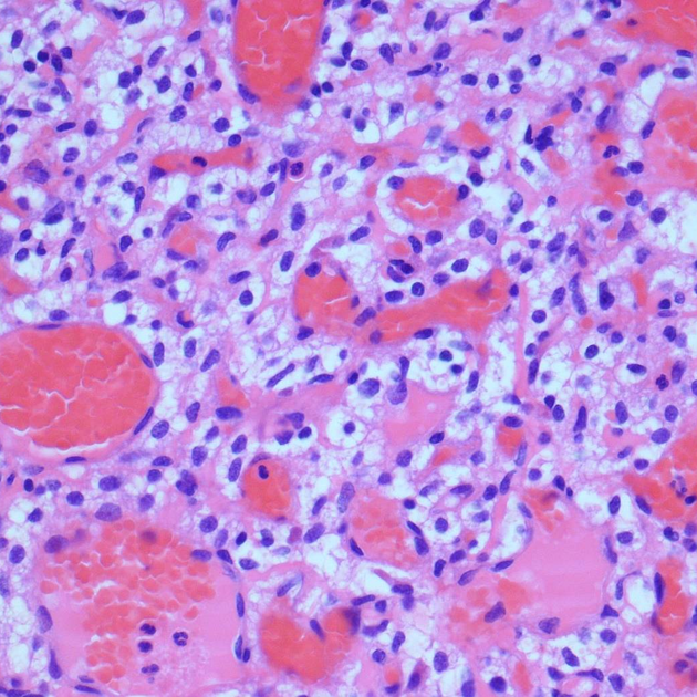
MICROSCOPIC DESCRIPTION: The sections show a moderately hypercellular intensely vascular tumor. Vascular channels vary from large caliber thin walled sinusoidal structures to small capillaries. The latter enclose lobules of cells with moderately pleomorphic round and oval hyperchromatic nuclei and clear cytoplasm. These are consistent with stromal cells. No mitotic figures or areas of necrosis are identified. The features are of capillary hemangioblastoma. Tumor is sharply demarcated from adjacent cerebellar parenchyma and focally is seen to be attached to overlying leptomeninges.
FINAL DIAGNOSIS: hemangioblastoma.
Case Discussion
This case illustrates the typical appearances of a hemangioblastoma: cystic mass with vividly enhancing mural nodule, with flow voids.


 Unable to process the form. Check for errors and try again.