
Presentation
Polycythemia for investigation.
Patient Data


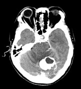
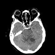
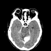
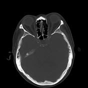
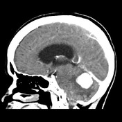
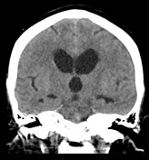
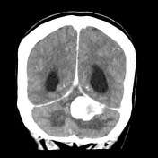

In the posterior fossa, abutting the inferior surface of the tentorium is a large vividly enhancing mass with a central region of cystic change.

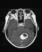
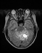
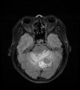
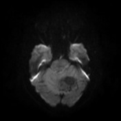
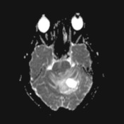
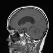
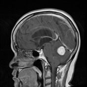
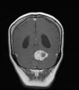
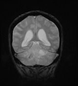
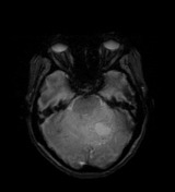
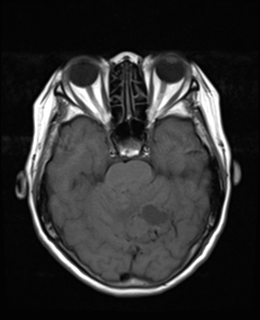
MRI confirms the presence of a vividly enhancing mass located peripherally in the left cerebellar hemisphere abutting the tentorium. There is surrounding high T2 signal consistent with edema. Best seen on T2 are prominent flow voids.
Features are almost certainly those of a hemangioblastoma.

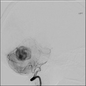
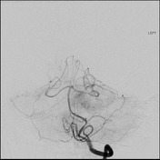
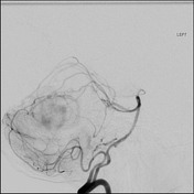
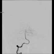
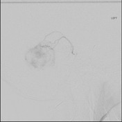
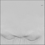
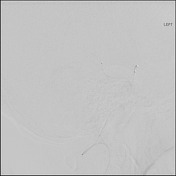
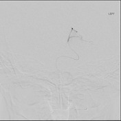
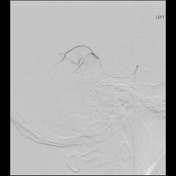
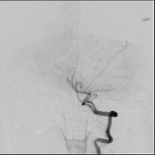
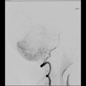
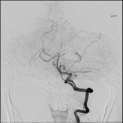
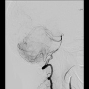
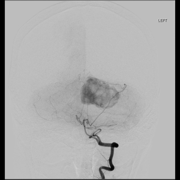
The tumor in the left superior cerebellar hemisphere near the vermis, is markedly hypervascular, with a rapid arteriovenous shunting very large veins at the dorsal and superior surface of the tumor. There are numerous arterial enlarged feeding arteries, predominantly from the left superior cerebellar artery, but with contribution from a small posterior cerebral artery branch, and left posterior inferior cerebellar artery, and to lesser extent anterior inferior cerebellar artery. A 5-French guiding catheter was placed in the left vertebral artery, and a SL team microcatheter navigated in to the left superior cerebellar artery, well beyond the brainstem, with selective, and embolization with PTA particles 150-250 micron. No neurologic complications, the procedure was tolerated well, there is good devascularisation of the tumor. Groin closure with six French AngioSeal device.
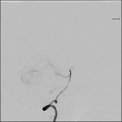
Conclusion: Marked decrease in tumor blush post pre operative embolization of hemangioblastoma
Case Discussion
The patient went on to have surgery.
Histology
MICROSCOPIC DESCRIPTION: The sections show scattered stromal cells in a background of many capillary-sized blood vessels. Some of the vessels contain foreign body material, consistent with preoperative embolization. The tumor cells have mildly enlarged nuclei with hyperchromasia and foamy vacuolated cytoplasm. Mitoses are inconspicuous. There is no necrosis. The features are those of hemangioblastoma. There is no evidence of malignancy.
FINAL DIAGNOSIS: Hemangioblastoma (WHO Grade I)


 Unable to process the form. Check for errors and try again.