
Presentation
Headache, known hypertension, on anticoagulants
Patient Data

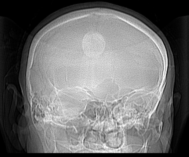
Frontal localizer (scout) image. 3 x 2.5 cm well-circumscribed circular opacity projected over the sagittal suture.
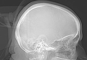
A lateral scout image shows that the radiopacity is external to the patient and would seem to represent their ponytail with hairband.
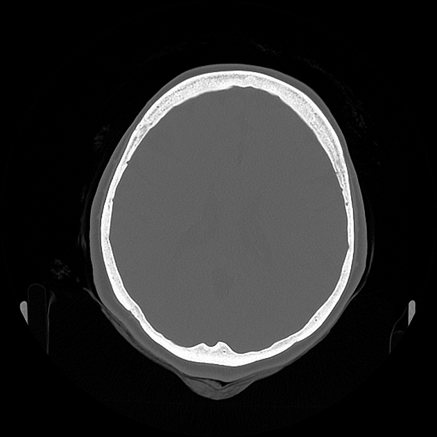
In case there is any doubt, the axial CT with bony reconstruction, unequivocally shows that the artifact is secondary to the pony tail behind the patient's head.
Case Discussion
Clothing, jewelry, hair accessories (etc.), can mimic pathology on all forms of imaging and therefore it is important to always be aware so that one is not caught out. Otherwise there is a risk of diagnosing pseudopathology. Hair-related artifacts are a common artifact on chest radiographs and head CTs.


 Unable to process the form. Check for errors and try again.