
Hemifacial microsomia
Updates to Case Attributes
Treacher-Collins syndromeThis case presents multiple radiological features of hemifacial microsomia, a condition characterised by hypoplasia of structures derived from the first and second branchial arches.
It is genetic disease characterisedassociated with extracranial anomalies in 55% of cases, as seen in this patient who has pentalogy of Fallot.
The main differential diagnosis is Treacher Collins syndrome, which is characterised by deformities ofbilateral involvement.
Another differential is Parry-Romberg syndrome, which is characterised by CNS and orbital manifestation and usually spares the ears, eyes, cheekbones, and chin and sometimes affect other systems as Heartear structures.
It is estimated to occur in one in 10,000 to one in 50,000 births.
Differential diagnosis include Miller syndrome and Nager syndrome but in both syndromes additional limb anomalies are seen.
Updates to Study Attributes
Facial asymmetry with underdeveloped right hemifacial bony and soft tissue structures as follows:
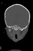
Osseous:
small-sized, hypoplastic right mandibular condyle with foreshortened and underdeveloped right mandibular ramus and mild subsequent ipsilateral deviation of the chin to the right side
small-sized right maxilla, maxillary antrum, and right pterygoid plate
Soft Tissue:
small-sized right masseter, temporalis, and pterygoid muscles, as well as the overlying subcutaneous soft tissue
absence of the right parotid gland
Auditory:
right microtia with aplastic external auditory canal and middle ear cleft, as well as mastoid air cells
Additional Finding:
right cerebellopontine angle arachnoid cyst
Image 1 ( destroy )
Image 1 ( destroy )
Image 1 ( update )
Image 1 CT (non-contrast) ( update )
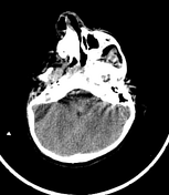
Image 48 CT (bone window) ( create )
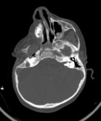
Image 61 CT (bone window) ( create )


 Unable to process the form. Check for errors and try again.