
Hepatic abscess ruptured into biliary tree with cholangitis and thrombosis of right hepatic vein
Presentation
Right upper quadrant pain, fever, and jaundice. Had cholecystectomy 20 years ago.
Patient Data


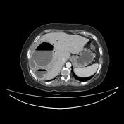


The CT scan demonstrates:
- large lobulated hypodense collection air-fluid level in the right hepatic lobe centered on the segments 7 and 8 with double target sign (the inner ring of abscess membrane shows early contrast enhancement and outer hypodense rim of parenchymal edema which only enhances on delayed phase)
- extensive linear branching gas within the liver most prominent in the left lobe and common bile duct (CBD) which is dilated with air-fluid in its distal portion. No distal CBD stone seen
- thrombosis of the right hepatic vein

Annotated image: the green arrow indicates most likely the site of rupture into the intrahepatic biliary tree.
Case Discussion
CT features most consistent with a large hepatic abscess ruptured into the intrahepatic biliary tree with infective cholangitis and thrombosis of the right hepatic vein in a patient presented with Charcot triad (fever, right upper quadrant abdominal pain, and jaundice). The possibility of ascending cholangitis complicated by hepatic abscess seems unlikely.
An urgent ultrasound/CT-guided drainage was requested, unfortunately, the patient died less than 24 hours after the CT scan due to septic shock.


 Unable to process the form. Check for errors and try again.