
Presentation
A liver lesion was incidentally detected in the right hepatic lobe on ultrasound.
Patient Data
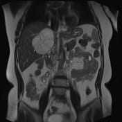

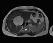
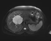
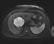
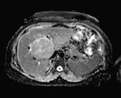
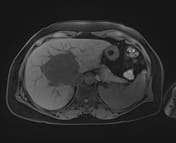
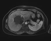
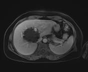
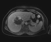
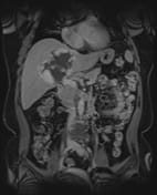
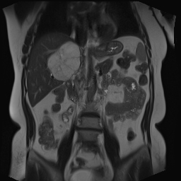
In the right hepatic parenchyma, adjacent to the inferior vena cava (mainly in subsegments V-VIII), there is a mass lesion measuring approximately 73 × 75 × 89 mm, with a lobulated margin and multiple internal septations. It exhibits low signal intensity on T1W and T1FS, high signal intensity on T2W and T2FS, diffusion restriction, and heterogeneous peripheral nodular enhancement, which progressively fills in a centripetal pattern on dynamic sequences.
Liver function and viral marker test results:
HCV Ab (automated Immunoassay): negative
HBsAg (automated Immunoassay): negative
quantitative AFP (Alpha-Fetoprotein) : 2.72 ng/mL (within normal range)
serum ALT (GPT) activity: 17.1 U/L (within normal range)
serum AST (GOT) activity: 19.3 U/L (within normal range)
Case Discussion
The imaging findings are consistent with hepatic hemangioma.
This is a benign lesion, and the patient is asymptomatic with blood test results within normal limits. The recommended approach for these patients is periodic follow-up ultrasound after 6 months or earlier if symptoms develop to confirm stability.


 Unable to process the form. Check for errors and try again.