
Presentation
Incidentally discovered hepatic lesions on ultrasound.
Patient Data
Age: 55 years
Gender: Male
From the case:
Hepatic hemangiomas


Download
Info
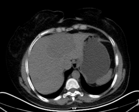
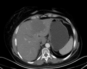
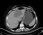
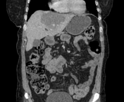
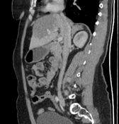
Two hypoattenuation lesions are seen in the left liver lobe as well as segment VII of the right lobe on non-enhanced CT images. Both of them show discontinuous, peripheral, nodular enhancement on arterial phase and progressive centripetal fill-in on portal and delayed phases.
Case Discussion
These typical findings are impressive of hepatic hemangiomas.


 Unable to process the form. Check for errors and try again.