
Presentation
Right flank pain. Non-contrast CT was performed to exclude urolithiasis.
Patient Data
Age: 40 years
Gender: Female
From the case:
Hepatic lipoma


Download
Info
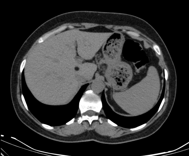
CT demonstrated a hypodense homogenous well defined rounded focal hepatic mass, measured 8 mm in diameter with a mean density -120-140 HU. Findings most consistent with a lipoma.
Case Discussion
Hepatic lipomas are asymptomatic extremely rare mesenchymal benign tumors. They are usually discovered incidentally and there is no risk of malignant transformation.


 Unable to process the form. Check for errors and try again.