
Patient Data


CT shows a hepatic nodule in segment VI that is hyperdense compared to steatotic liver parenchyma. After contrast administration is noted a progressive peripheral enhancement that is centripetal and has a delayed homogenization.
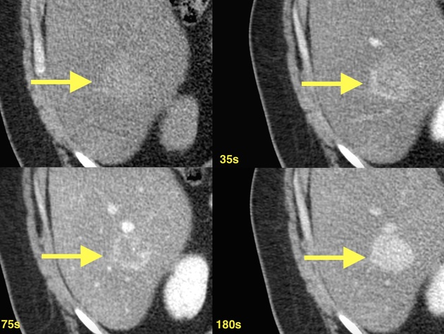
CT shows a hepatic nodule in segment VI that is hyperdense compared to steatotic liver parenchyma. After contrast administration is noted a progressive peripheral enhancement that is centripetal and has a delayed homogenization.
Case Discussion
Diffuse hepatic steatosis is a common imaging finding, and can lead to difficulties assessing the liver appearances, specially on ultrasound exam.
Hepatic hemangioma is a benign non-neoplastic hypervascular liver lesions. They are frequently diagnosed as an incidental findings on imaging and most patients are asymptomatic.


 Unable to process the form. Check for errors and try again.