
Presentation
Incidental lesion on ultrasound of the liver (not shown).
Patient Data
Age: 70 years
Gender: Male


Download
Info
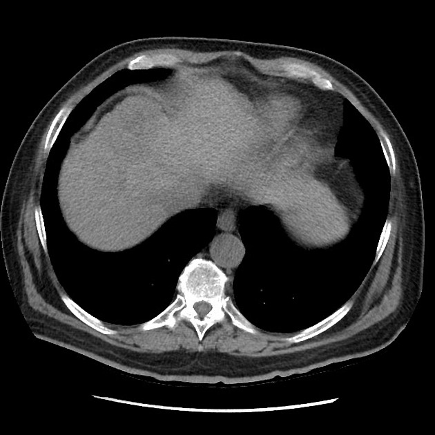
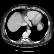
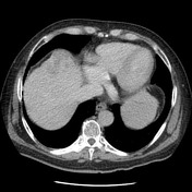
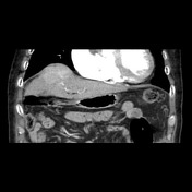
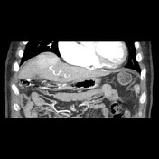
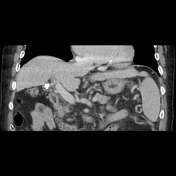
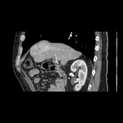
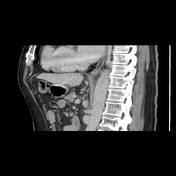
The liver is cirrhotic and demonstrates a large (6cm) hypervascular lesion in the segment IVA that shows washout on the subsequent portal venous and delayed phases. Minor shunting of the contrast to the hepatic vein is demonstrated during arterial phase. The features are consistent with hepatocellular carcinoma. No other focal hepatic lesion is identified. The portal and hepatic veins are patent.
Case Discussion
Typical imaging features of HCC in a cirrhotic patient.


 Unable to process the form. Check for errors and try again.