
Presentation
Right-sided abdominal pain referred to the back, nausea, sometimes vomiting. History of cholecystectomy. ALP 835 IU/L (N 44 to 147 IU/L), AST 79 (N 10 to 36 U/L), ALT 59 (N 4 to 36 U/L)
Patient Data
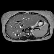

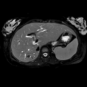
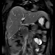
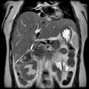


Multiple T2 signal void structures (calculi) are seen in the main right and left intrahepatic bile ducts proximal to common hepatic duct with moderately dilated intrahepatic biliary radicles on both hepatic lobes.
Choledochojejunostomy is noted with a stent seen.
The main pancreatic duct is not dilated. Cholecystectomy. Beaver tail of the liver (normal variant).
Case Discussion
A case of hepatolithiasis in a post-cholecystectomy patient. Patients may develop common hepatic duct strictures, that predisposed to intrahepatic biliary calculus formation.
Patients with primary hepatolithiasis usually present in 5th and 6th decades. Patients presenting in the 3rd decade, as this patient, are commonly to have autoimmune hepatitis, sclerosing cholangitis or a history of open cholecystectomy 1.


 Unable to process the form. Check for errors and try again.