
Presentation
Multiple cutaneous and tongue telangiectasias with recurrent epistaxis.
Patient Data
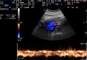

On ultrasound examination, the gallbladder appeared free from lithiasis, while in B-mode were identifiable tortuousness of the intrahepatic branches of the liver artery, dilation of the liver artery with accelerated flows and arterial malformations in the liver parenchyma. The ultrasound features are suggestive for HHT.
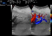
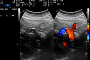
Images 1-2: Arteriovenous malformation that connects left portal branch and left suprahepatic vein.
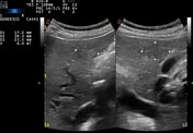
Images 3-4: Tortuousness of the hepatic artery.
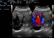
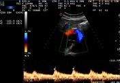
Images 5-6: Enlarged hepatic artery with accelerated flow.
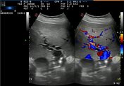
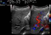
Images 7-8: Focal liver lesion consisting of hepatic telangiectasia.
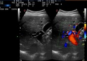
Image 9: Portal vein dilatation with flow acceleration.
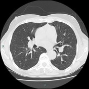
CT chest & abdomen C+
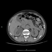

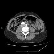
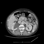

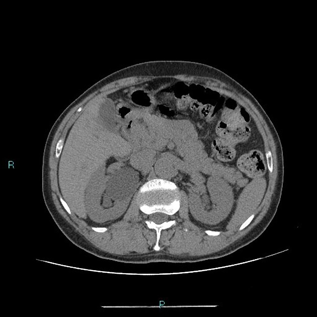
During the arterial phases, liver shows heterogeneous perfusion pattern with millimetric hypervascular images disseminated throughout the hepatic parenchyma, referring to telangiectases and arteriovenous shunts that are no longer evident during the venous phase. The early venous drainage with the simultaneous opacification of the dilated hepatic veins and the hepatic artery is evident during arterial phase. These finds are no longer evident during the venous phase. Hepatic artery enlargement: 10 mm (i.e. diameter > 6.5 mm) and portal vein enlargement: 17 mm (i.e. diameter > 13 mm). The liver and splenic size are normal. Some telangiectasias are present in the pancreas. There’s modest bile ducts dilatation.
Case Discussion
Hepatic abnormalities include enlarged hepatic arteries, hepatic artery aneurysms, telangiectasia and arteriovenous fistulae. Hereditary hemorrhagic telangiectasia (HHT) is an autosomal dominant disorder. In the arterial phase, the liver shows a heterogeneous mosaic-like perfusion pattern due to multiple arteriovenous shunts that show different attenuations and telangiectasias.
Telangiectasias are rounded hypervascular nodules more frequent in the periphery. The vascular supply of the bile ducts depends on the branches of the hepatic artery, so shunts by subtracting the arterial flow can cause ischemic cholangitis. The bile ducts dilatation it may also due to the compression of enlarged vascular structures.
Case courtesy: Dr. Luca Giampaolo


 Unable to process the form. Check for errors and try again.