
Hereditary multiple exostoses of the pelvis and proximal femora
Presentation
Bilateral coxalgia.
Patient Data
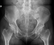
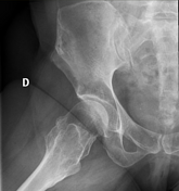
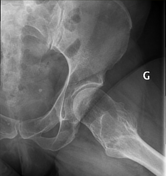

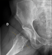
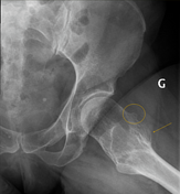
Bilateral coxa valga, short and widened femoral neck, superior pubic ramus widening, multiple small osteochondromas of the femoral neck, and intertrochanteric crest.
Mild coxarthrosis with preserved head sphericity.
Remodeling of the tuberculum of the right iliac crest related to previous surgical excision of a lesion, presumably related to the malignant transformation of an osteochondroma.
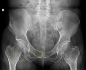
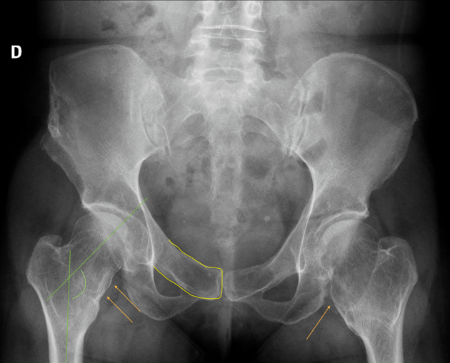
Yellow lines outline widened superior pubic ramus.
Green lines depict coxa valga deformity of the hip where there's a widened angle between the femoral head-neck and its shaft.
Orange arrows and circle point to the osteochondromas that superimpose on the frontal view.
Case Discussion
Almost pathognomonic findings of multiple hereditary exostoses of the pelvis and hip.
Here, bilateral and symmetric involvement with many small, sessile or pedunculated bone growths extending outwards from the femoral neck and intertrochanteric surfaces.
There's a risk of malignant transformation into chondrosarcoma. This may explain the surgical excision at the right iliac crest (however, no pathological report in the patient's medical record to confirm it).


 Unable to process the form. Check for errors and try again.