
Presentation
Intermittent left loin pain and non-visible hematuria. Cystoscopy and ultrasound normal.
Patient Data


Previous bowel surgery with colonic suture line. No renal masses or renal tract stones.
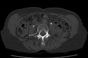
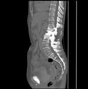

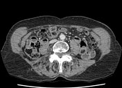
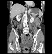
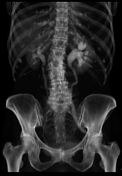
The left renal pelvis and ureter are distended, secondary to entrapment of the distal ureter within a left sciatic hernia (trapped within the greater sciatic foramen). This has an infrapiriformis location. There is no herniation of bowel. There is blunting of the calyces of the left kidney, but overall renal size is not reduced when compared to the right kidney.


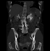
The 3D reconstructions from the delayed phase CT dataset show the abnormal path of the left ureter into the left greater sciatic foramen. This has been described as the curlicue ureter sign.
The 3D reconstruction from the delayed phase CT dataset show the abnormal path of the left ureter into the left greater sciatic foramen. This has been described as the curlicue ureter sign.
Case Discussion
The distal left ureter is trapped within a ureterosciatic hernia causing upstream obstruction. It enters the greater sciatic foramen, inferior to the piriformis muscle. A conservative management path was followed.


 Unable to process the form. Check for errors and try again.