
Presentation
Hip pain.
Patient Data

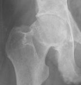
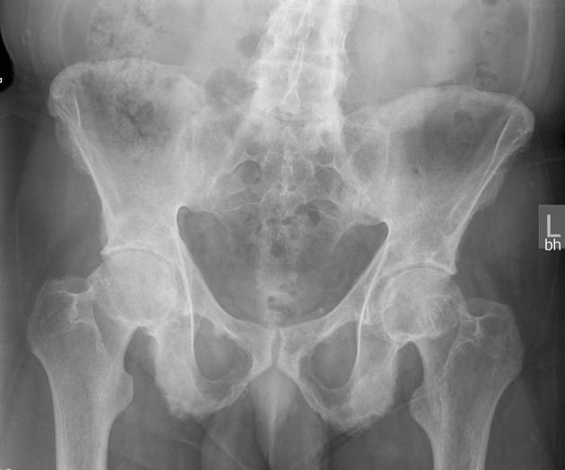
Evidence of concentric narrowing of the hip joints, femoral osteophytosis and subchondral sclerosis. The hip joints are asymmetrically affected, greater on the right. There is ossification of the ligamentous origins in particular at the ischial tuberosities giving a furry appearance. Near complete fusion of the sacroiliac joints with erosive changes at the pubic symphysis.

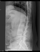

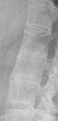
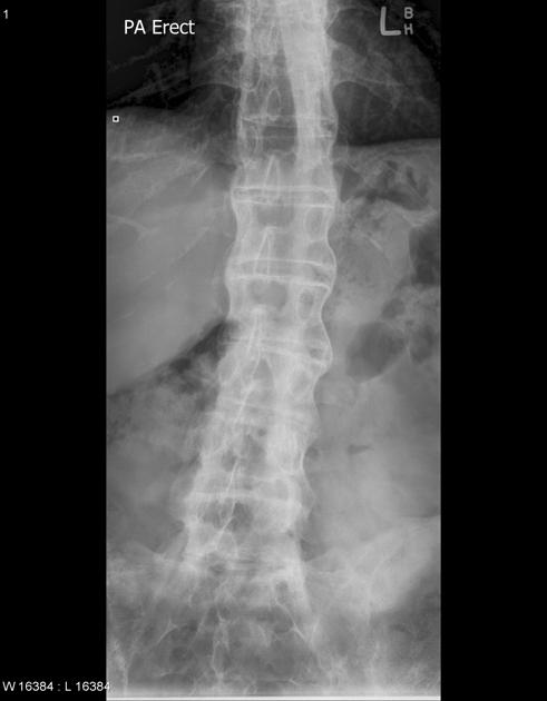
Syndesmophytes are present with "squaring" of the thoracolumbar spine and fusion of the mid lumbar vertebrae.
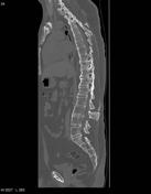

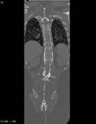

1. Sagittal and 2. coronal CT images shows syndesmophytes in the outer fiber of annulus fibrosis and osteitis at the anterior corners of the vertebral bodies. The bone is osteopenic with long column fusion of vertebral bodies and facet joints with skipped intervening regions.
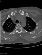
3. Axial CT images shows erosive changes in the sternoclavicular joints.


 Unable to process the form. Check for errors and try again.