
Presentation
Something protruding in upper thigh, long standing history.
Patient Data
Age: 65 years
Gender: Female
From the case:
Hip prosthesis

Download
Info
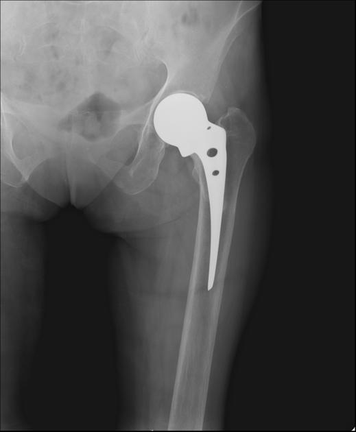
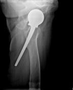
The frontal view of the left hip appears relatively normal, demonstrating a left hip prosthesis.
Dislocated stem of prosthesis is however seen clearly on the lateral view.
Case Discussion
Prosthesis was inserted 16 years previously. Presently complaint of 'pricking' type pain in the upper thigh. No difficulty in walking at present.
This case explains why two radiograph at orthogonal planes are required. If a second view is not taken then important pathology may be missed.


 Unable to process the form. Check for errors and try again.