
Presentation
Axial load to hypothenar eminence after falling onto golf club handle
Patient Data


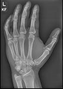
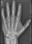

Standard lateral, oblique and frontal radiographs were acquired. Special lateral view with thumb abduction and radial deviation of the hand was also performed due to high clinical suspicion of hamate fracture.
Fracture through the hook of the hamate with ~2mm of diastasis between the fracture fragments, best seen on lateral and thumb abduction views.


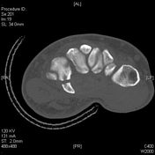
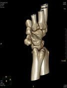
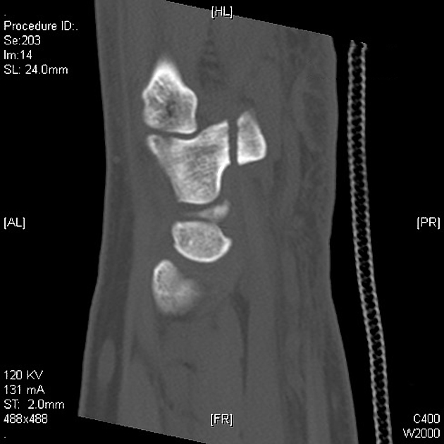
Acute transverse fracture through the base of the hook of hamate, with minor separation of the fragments by 2mm. Features in keeping with a Milch Type I hamate fracture (subtype III (base of hook)).
Case Discussion
Case contributed by Dr. James Willard-Jones
Hook of Hamate fractures are uncommon, and account for <4% of carpal fractures. These fractures are often missed, causing delayed diagnosis and treatment. It is particularly important to identify these fractures as they are at risk of non-union. Hamate fractures are organized using the Milch Classification, which divides them into fractures of the Hook (type I) and Body (Type II). Hook fractures can be subsequently subdivided into three types: subtype 1 (distal tip), subtype 2 (body of hook) and subtype 3 (base of hook). The most common is subtype 3, as in the case above, which accounts for 75% of hook of hamate fractures1.
While readily identifiable on CT imaging, the initial modality of plain radiographs can often obscure the diagnosis. As such, if there is a high clinical suspicion for a hook fracture then additional views are recommended in addition to the standard 3-views. These include (a) lateral view with thumb abduction and radial deviation, (b) supinated oblique and (c) carpal tunnel views.


 Unable to process the form. Check for errors and try again.