
Presentation
G2P1. Previous inconclusive scan at 5 weeks gestation presents with hyperemesis. ? viability.
Patient Data
Age: 40 years
Gender: Female
From the case:
Hydatidiform mole
Download
Info
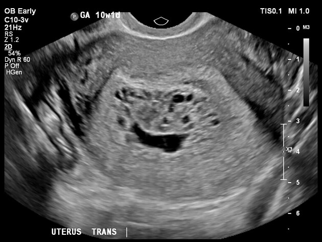
- distended uterine cavity by an enlarged heterogenous placenta showing multi-cystic 'bunch of grapes' appearance
- small volume anechoic fluid, which may represent either an irregular empty gestational sac, which is being compressed and displaced by the heterogenous mass or simple fluid within the cavity
- no definite fetal parts
- the mass demonstrates increased vascularity on Doppler assessment
- no myometrial invasion
- right ovarian corpus luteum
- no pelvic free fluid
Case Discussion
Hydatidiform moles are within the spectrum of gestational trophoblastic disease, which results in abnormal proliferation of trophoblastic tissue.
This patient's beta HCG was very high relative to the expected conception date.


 Unable to process the form. Check for errors and try again.