
Hypertrophic olivary degeneration (bilateral) following midbrain hemorrhage
Presentation
Sudden onset diplopia and weakness.
Patient Data
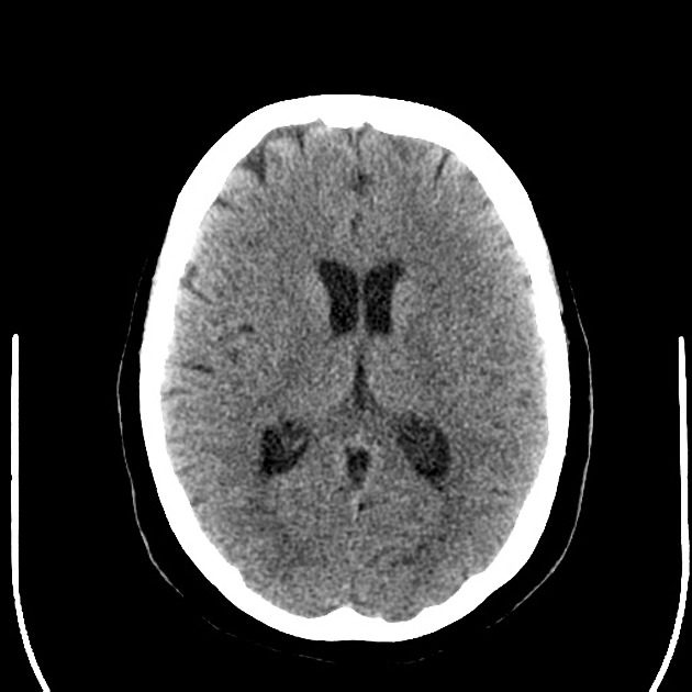
There is a right tectal hematoma extending into the 4th ventricle. A small rim of edema surrounds the hematoma and there is compression of the right ambient and quadrigeminal plate cisterns. There is very mild dilatation of the lateral ventricle temporal horns. The remainder of the brain is unremarkable.
Bilateral paranasal sinus mucosal thickening and left sphenoid sinus mucus retention cyst noted.
MRI 6 months later


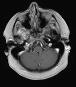
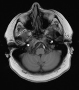
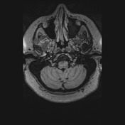
The region of previous hemorrhage within the right side of the midbrain, extending inferiorly to the upper dorsal pons, just lateral and above the facial colliculus is again demonstrated, with no underlying lesion evident (no enhancing mass lesion, or abnormal vessels).
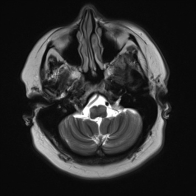
Since the hemorrhage, bilateral T2 hyper-intensity is seen involving the inferior olivary nuclei more so on the left and both are slightly swollen.
Conclusion: Features are pathognomonic of hypertrophic olivary degeneration, due to interruption of the right central tegmental tract, and crossing dentatorubral tract, accounting the bilateral involvement. Clinical correlation with palatal myoclonus would be useful.


 Unable to process the form. Check for errors and try again.