
Presentation
Trauma to the left side of neck with hoarseness of voice.
Patient Data
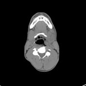

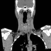
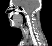
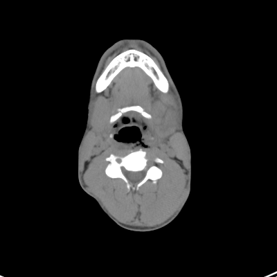
Focal defect in the left posterolateral wall of the hypopharynx with a related air-filled tract extending posterolaterally. Multiple related air loculi are present in the pharyngeal wall, left para-pharyngeal space, and prevertebral space, as well as surrounding the cervical oesophagus, extending to the thoracic inlet (emphysema). A small focal air outpouching is also noted at the left lateral wall of the oropharynx.
Soft tissue thickening and irregular mucosal outline are observed at the supraglottic larynx.
Enlarged left submandibular gland with heterogeneous density, showing branching hypodense lines denoting laceration. Associated surrounding soft tissue oedema, blurring of fat planes, and thickening of the skin in the left submandibular and submental regions are also noted.
Asymmetrical vocal cords.
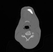
Fractures of spinous processes of C6 and C7 vertebrae.
Case Discussion
Hypopharyngeal rupture due to blunt neck trauma is an extremely rare clinical entity. Early diagnosis and appropriate management are crucial, as an overlooked injury can lead to catastrophic consequences 1.
In this case, neck emphysema should prompt a search for the source. A thorough examination of the airway reveals a defect in the left posterolateral wall of the hypopharynx, indicating that the hypopharyngeal injury is the source of the air 1.
Submandibular gland laceration is also rare due to its protection by the overlying mandible 2.
As in our case, hypopharyngeal rupture usually occurs at the level of Killian's dehiscence as this anatomical landmark represents the parietal weakness of the hypopharynx with retraction of the muscle fibres and only persistence of the mucosal and serosal layers 3. The Killian's dehiscence is between the inferior pharyngeal constrictor and cricopharyngeal muscles 3.
The mechanism of hypopharyngeal rupture occurs by an upper airway closure due to compression at the level of the hyoid bone concurrently with expiration. The generated pressure leads to hypopharyngeal rupture 3.
Radiographic features include the presence of hypopharyngeal wall defect with air leak through the defect into the retropharyngeal/prevertebral spaces. Gatsrograffin swallow-enhanced CT scan is the best diagnostic tool for detecting the location and extension of the tear 3.
Management might be conservative or surgical according to the size and the location of the tear as well as the clinical condition of the patient 3 .


 Unable to process the form. Check for errors and try again.