
Presentation
VF arrest.
Patient Data


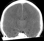
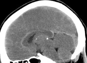
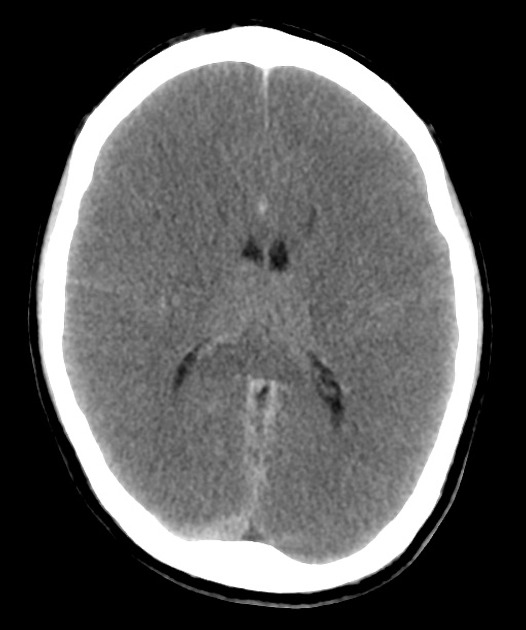
Decreased grey-white differentiation at the vertex with complete loss of deep grey matter at the caudate heads extending into the lentiform nucleus. The thalami are spared. There is effacement of convexity sulci and partial effacement of the ventricles and basal cisterns. These findings are in keeping with gross cerebral edema. No intra- or extra-axial collection or hemorrhage demonstrated. Apparent hyperdensity of the basal cisterns and tentorium cerebelli is in keeping with pseudosubarachnoid sign. Partial opacification of the sphenoid and ethmoid sinuses.
Conclusion:
Findings in keeping with global ischemic brain injury.
Case Discussion
This case demonstrates multiple features of global hypoxic brain injury including pseudosubarachnoid hemorrhage.


 Unable to process the form. Check for errors and try again.