
Presentation
Headache
Patient Data
Age: 50 years
Gender: Female


Show annotations
Download
Info
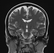
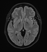
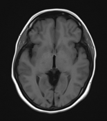
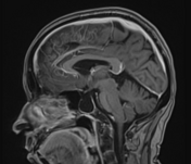
Mildly enlarged and empty sella.
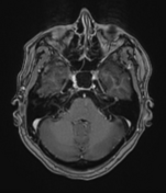
Mildly enlarged bilateral Meckel cave CSF space.
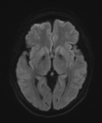
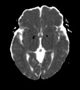
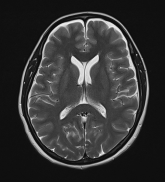
Mild focal flattening of posterior sclera on either side.
Prominent optic nerve sheath CSF space.
The optic nerve tortuosity is on either side.
There is no evidence of transverse sinus stenosis on either side.
Case Discussion
On fundoscopic examination, there was bilateral papilledema and the CSF opening pressure was 30 cm H2O.
So the MRI findings, fundoscopy findings and high CSF opening pressure are suggestive of idiopathic intracranial hypertension.
Case co-author: Dr Abhishek Kumar, Neurologist, Medanta Hospital, Patna, India.


 Unable to process the form. Check for errors and try again.