
Presentation
The patient presented with headache.
Patient Data


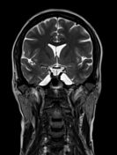
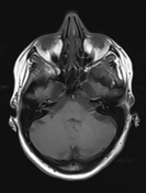
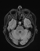
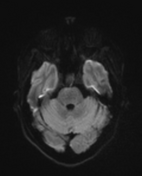
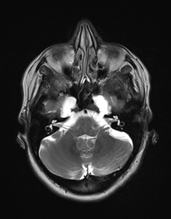
Bilateral dilatation of Meckel's cave (axial, coronal) with posterolateral extension to petrous apex on both sides suggesting bilateral petrous apex meningocele (axial, coronal).
Small meningocele at the jugular foramen on both sides (axial, coronal).
Small hypoglossal canal meningocele on the right side (axial, coronal).
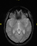
Dilated tortuous optic nerve sheath diameter (ONSD) on both sides (axial, sagittal).
Prominent oculomotor cistern on both sides.
Small arachnoid granulation at the right transverse sinus (axial, coronal).
Prominent perivascular spaces.
No intracranial space occupying lesions or definite signs of dural sinus thrombosis.
Case Discussion
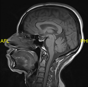
The radiological features of enlarged arachnoid outpouching (enlarged Meckel's caves, partial empty sella, arachnoid pits, skull base meningoceles, prominent oculomotor cisterns, perivascular spaces) and dilated tortuous optic nerve sheath diameter are highly suggest idiopathic intracranial hypertension.


 Unable to process the form. Check for errors and try again.